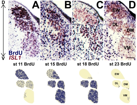
Fig. 3.
Inside-out gradient of OMC neurogenesis. Chick midbrains were processed at E12 for BrdU immunohistochemistry (blue) and ISL1 gene expression (brown/pink) after BrdU delivery to st 8-23 embryos. Coronal sections (top) and corresponding BrdU chartings (bottom) illustrate the dorsal-to-ventral (D↔V) progression of OMC neurogenesis. Panels illustrate the left OMC, with the midline to the right. OMC subnuclei are identified in D. (A) Heavy BrdU labeling in the EW nucleus indicates that neurogenesis of visceral oculomotor neurons precedes that of somatic oculomotor neurons. (B) Extensive BrdU labeling is seen throughout the OMC. (C,D) Neurogenesis is nearly complete for the EW nucleus by st 18 (C) and for the somatic motoneurons by st 23 (D). Scale bar: 0.1 mm.

