
Figure 1.
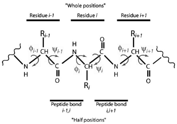
Diagram of protein backbone, including φ and ψ angles, whole positions, and half positions. At the ith residue the φ angle describes the torsion around the bond Ni–Cαi, measuring the angle between the Ci−1–Ni and the Cαi–Ci bonds, whereas the ψ angle describes the torsion around the bond Cαi–Ci, measuring the angle between the Ni–Cαi and the Ci–Ni+1 bonds. (In the graphic, CH represents a Cα atom and the attached hydrogen atom.) The torsion angle pair (φ, ψ) on either side of a residue R is considered a whole position. Three such pairs are shown. The torsion angle pair (ψ, φ) on either side of a peptide bond, between two residues, is considered a half position. Two such pairs are shown.