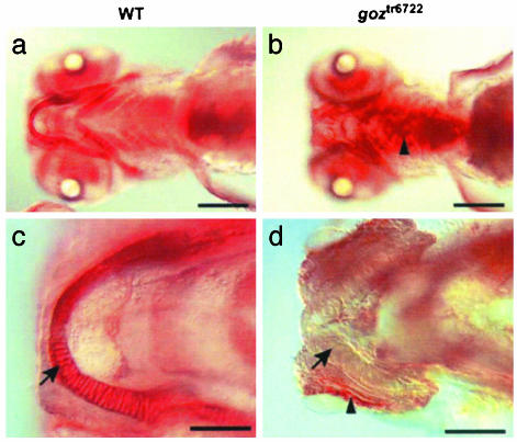
Fig. 2.
Proteoglycan phenotypes in goz. Wheat germ agglutinin staining of matrix proteoglycans in wild-type (WT) sibling larvae (a and c) and goz larvae (b and d) at 5 dpf. (a and b) Ventral view. (c and d) Enlargement of Meckel's cartilage. Proteoglycans are homogeneously stained in the cartilage matrix of sibling larvae (a and c). This staining is absent in the cartilage matrix of goz (arrow in d) where proteoglycans seem to accumulate ectopically (arrowheads in b and d). (Scale bars are 200 μm in a and b and 50 μm in c and d.)

