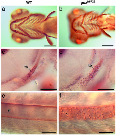
Fig. 3.
Col II defects in goz. Immunohistological staining of Col II in wild-type (WT) sibling larvae (a, c, and e) and goz larvae (b, d, and f) is shown. (a and b) Ventral view of the head 5 dpf. (c and d) Enlargement of the trabeculae (tb) of the neurocranium (lateral view) at 2 dpf. (e and f) Enlargement of the notochord (n) in the tail at 2 dpf. Cartilage matrix is homogeneously stained in sibling larvae (a, c, and e), whereas abnormal protein aggregates can be seen in the cartilage matrix and around the notochord in goz larvae (d and f). (Scale bars are 200 μm in a and b and 50 μm in c–f.)

