
Figure 1.
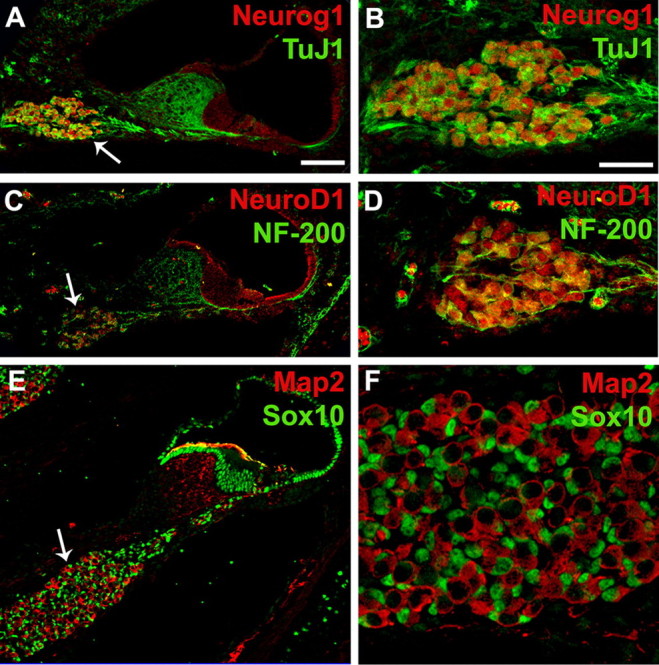
Neurog1 and NeuroD1 are expressed in spiral ganglion neurons. A, Cross-section through the cochlea at P0 illustrating expression of Neurog1 (red) and ΤuJ1 (green) in the spiral ganglion (arrow). B, High-magnification view of the spiral ganglion labeled as in A. Note that TuJ1 and Neurog1 are coexpressed in neuronal cells. C, Cross-section as in A illustrating expression of NeuroD1 (red) and NF-200 (green) in the spiral ganglion (arrow). D, High-magnification view of the spiral ganglion labeled as in C. NeuroD1 and NF-200 are colocalized in spiral ganglion neurons. E, Cross-section as in A, illustrating expression of Map2 (red) and Sox10 (green). Map2 is expressed in neurons while Sox10 is expressed in nuclei of spiral ganglion glia (arrow) and nonsensory cells in the cochlear duct. F, High-magnification view of the spiral ganglion labeled as in E. Large Map2-positive neurons are surrounded by smaller Sox10-positive glial cells. Scale bars: A (for A, C, E) 50 μm; B (for B, D, F) 20 μm.