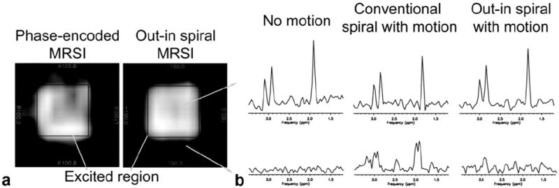
FIG. 4.

a: Reconstructed water phantom images from phase-encoded MRSI (left) and out–in spiral (right) acquisitions in the presence of motion similar to respiration. Due to motion, normal phase-encoded MRSI results in artifacts appearing outside the excited PRESS box. This is reduced with an out–in spiral acquisition. Motion was induced in the R–L direction. b: Representative metabolite spectra from a spectroscopy phantom experiment using conventional and out–in spiral. The top row illustrates spectra obtained from within the excited region. The reconstructed data using an out–in spiral has increased SNR and reduced artifacts compared to the conventional spiral readout.
