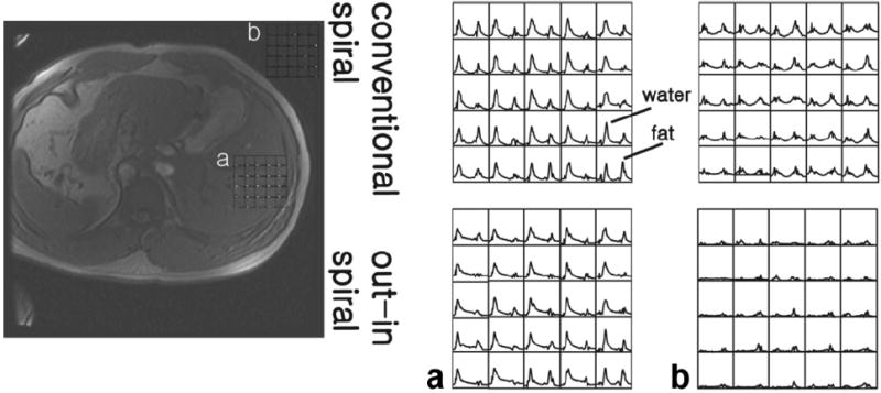
FIG. 5.

Water and fat spectra obtained from an in vivo study of a healthy adult volunteer. Selected spectra from 32 × 32 acquisition using conventional and out–in spirals are shown from within the liver (a) and regions outside of the body (b). Due to motion effects, data reconstruction with the conventional spiral resulted in spectral artifacts appearing outside of the body (b). This is reduced using an out–in spiral trajectory. Overall, out–in spirals reduce effects due to motion allowing spatial variations of water and fat content to be readily observed.
