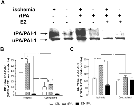
Fig. 3.
A, representative Western blot of PAI-1 in control, rtPA, and E2 + rtPA group at 24 h after transient focal MCAO by suture. B, densitometric analysis of PAI-1 in complex with tPA in control, rtPA, and E2 + rtPA group. A significant increase of PAI-1 in complex with tPA was demonstrated, which was further enhanced by rtPA treatment. E2 treatment attenuated PAI-1 in complex of tPA in both ischemic and contralateral hemisphere. C, densitometric analysis of PAI-1 in complex with uPA in control, rtPA, and E2 + rtPA group. rtPA significantly increased PAI-1 in complex with uPA in the ischemic hemisphere, which was attenuated by E2 treatment. ∗, p < 0.05.

