Fig. 2.
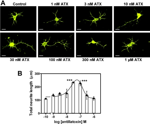
Effect of ATX on neurite outgrowth. A, representative images of DiI-loaded immature cerebrocortical neurons at 24 h after plating (scale bar, 10 μm). Various concentrations of ATX were added to the culture medium at 3 h after plating. Depicted neurons were visualized by diolistic loading with DiI. B, quantification of concentration-response effects of ATX on neurite outgrowth at 24 h after plating. ATX-enhanced neurite outgrowth displayed a hormetic concentration-response relationship with maximal enhancement seen at 30 to 100 nM ATX. Quantification of total neurite length was performed with Image Pro Plus. The experiment was performed twice, and each point represents the mean value derived from analysis of 25 to 30 neurons. ***, p < 0.001, unpaired t test.

