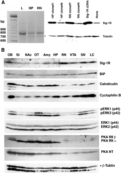
Fig. 4.
Brain expression and distribution of σ-1 receptors (Sig-1Rs), ER chaperones, PKA, and ERK. A, expression of Sig-1R transcripts in the rat brain. Left, the result of RT-PCR using total RNAs from the liver (L), hippocampus (HP), and red nucleus (RN). One microgram of rat liver total RNA or 2 μg of rat brain total RNA was applied to RT-PCR. ∗, the 5-methyltetrahydrofolate-homocysteine methyltransferase gene nonspecifically amplified by the Sig-1R primers. All the PCR products were identified by gene sequencing. Western blotting of Sig-1Rs (right) verified the cloned HP or RN cDNA expressing Sig-1Rs in the mammalian cells. Total cell lysates (15 μg/lane) were prepared from CHO cells transfected with PCR products cloned in the pcDNA3.1 vector. HP clone 6 and RN clone 4 serve as negative controls containing the nonfunctional gene. Sig-1R cDNA is a positive vector containing the Sig-1R cDNA previously cloned (Hayashi and Su, 2001). B, brain distribution of ER chaperones and kinases. See Materials and Methods for details.

