Figure 4.

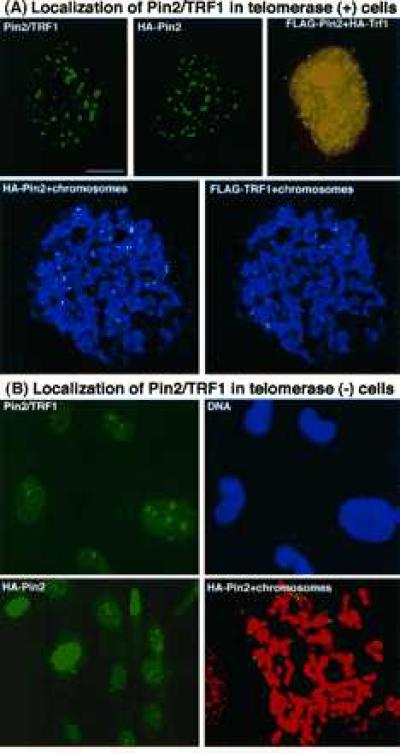
Differential localization of Pin2 is in telomerase-positive and -negative cells. (A) Localization of Pin2 protein in telomerase-positive HeLa cells. Pin2/TRF1, localization of the endogenous Pin2/TRF1 detected by immunostaining with anti-Pin2 antibodies; HA-Pin2, localization of the ectopically expressed Pin2 detected by staining with the 12CA5 mAb; HA-Pin2 and FLAG-TRF1, colocalization of expressed Pin2 and TRF1 detected by staining doubly transfected cells with 12CA5 and M2 mAbs, with the yellow image indicating a colocalization produced by superimposing the green Pin2 and the red TRF1 images; HA-Pin2/FLAG-TRF1+chromosomes, colocalization of expressed Pin2 and TRF1 evenly at telomeres detected by staining mitotic chromosomes from doubly transfected cells with 12CA5 and M2 mAbs. Green, HA-Pin2; red, FLAG-TRF1; blue, chromosomes. (B) Localization of Pin2 protein in telomerase-negative MDAH087 cells. Pin2/TRF1, localization of the endogenous Pin2/TRF1 detected by staining with anti-Pin2 antibodies (green) and the DNA dye Hoechst (blue); HA-Pin2, localization of the ectopically expressed Pin2 detected by staining a single stably HA-Pin2-expressing cell line (clone 6) with the 12CA5 mAb; HA-Pin2+chromosomes, localization of expressed Pin2 at telomeres detected by staining clone 6 mitotic chromosomes with the 12CA5 mAb. Green, HA-Pin2; red, chromosomes.
