
Figure 8.
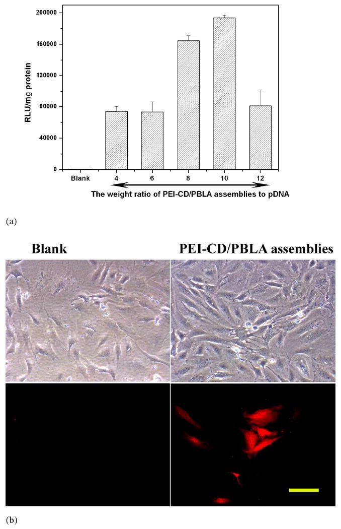
(a) In vitro transfection of the luciferase gene into osteoblast cells by PEI-CD/PBLA nano-assemblies. Luciferase activity (RLU) was quantified using luminometer 48 h after cells were transfected. Results were normalized to total cell protein. All samples were run in triplicate and on three or more separate occasions. (b) The transfected cells were viewed by a fluorescence microscope (Nikon Eclipse 50i microscope) 48 h after transfection. Excitation was performed with green light. Polyplexes with the weight ratio of PEI-CD/PBLA to pDNA of 8 were employed. The scale bar represents 100 μm.