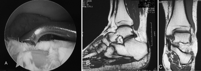
Fig. 1A–C.
(A) An ankle arthroscopy image shows a large, full-thickness chondral lesion down to subchondral bone. (B) A sagittal MR image for this patient shows no evidence of the lesion with no change in the bone marrow signal. (C) A coronal MR image for the same patient similarly shows no evidence of a lesion.

