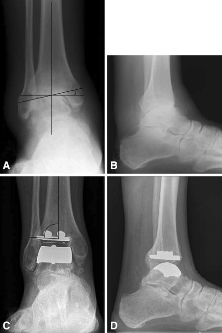
Fig. 1A–D.
(A–B) Preoperative and (C–D) 4-year postoperative AP and lateral weight bearing radiographic views of ankle arthrosis treated via the STAR prosthesis are shown. (A) Preoperative coronal deformity (11°) was measured with the angle of a line drawn along the superior talar dome relative to a line perpendicular to the mechanical axis of the tibia. (C) Measurement of varus or valgus of the prosthesis (88°) is shown. (D) Lateral radiograph at 4 years is shown.

