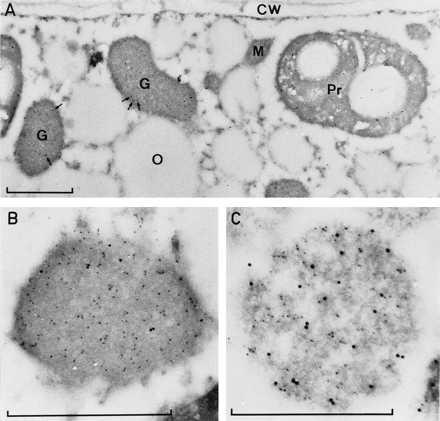
Figure 2.

Electron micrographs of aldehyde-fixed cells from 3-day-old watermelon cotyledons (A and B) or isolated glyoxysomes (C). The thin sections were treated with a-hsp70 antibodies and these visualized by 15 nm gold-conjugated goat anti-mouse antibodies; they were at the same time treated with a-gMDH antibodies that were visualized with 5 nm gold-conjugated goat anti-rabbit antibodies. Arrows indicate 15 nm gold particles in glyoxysomes (A). G, glyoxysome; Pr, proplastid; M, mitochondria; O, oil body; CW, cell wall. Bars = 0.5 μm.
