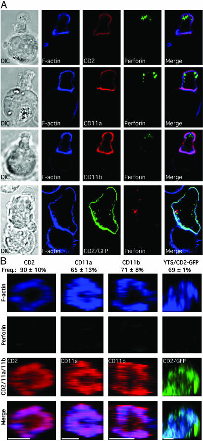
Fig. 5.
Perforin, but not cell-surface receptor or F-actin accumulation at the activating NKIS, depends on microtubule function. (A) Ex vivo NK cells or YTS-CD2/GFP cells treated with colchicine were conjugated with K562 or 721.221 cells, respectively. Layout, specific molecules evaluated, and colors displayed are the same as for Fig. 1. (B) Synapses from colchicine-treated ex vivo NK or YTS-CD2/GFP were analyzed throughout their volume and reconstructed in the z, x plane. The colors, specific molecules evaluated, and layout are the same as in Fig. 2. (Scale bars, 5 μm).

