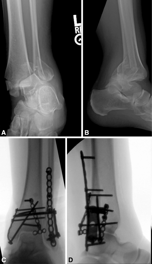
Fig. 3A–D .
(A) Anteroposterior and (B) lateral radiographs show a fracture-dislocation with complete dislocation and trimalleolar fractures. This is a typical example of a patient treated with combined fixation. (C) Anteroposterior and (D) lateral radiographs show combined posterior malleolar and syndesmotic screw fixation.

