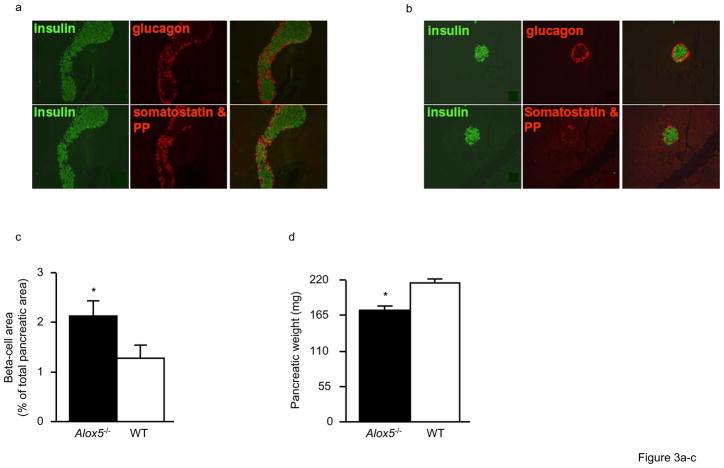
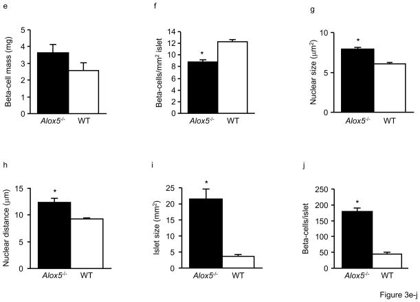
Figure 3. Morphology and histology of the pancreas in Alox5−/− and WT mice.
Double immunostaining of representative pancreatic sections (250X) for insulin (green), glucagon, somatostatin, and pancreatic polypeptide (PP) shows normal islet morphology and cellularity in both Alox5−/− (a) and WT (b) mice. The right hand panel for each genotype group shows overlaid immunostaining images. Alox5−/− mice have increased beta-cell area as a percentage of total pancreatic area (c) but decreased pancreatic weight (d). Beta-cell mass is not significantly different (e). The number of beta-cells counted per mm2 islet is decreased in Alox5−/− mice (f), suggesting the presence of hypertrophic cells. This supported by the increased nuclear size (g) and distance between nuclei of adjacent beta-cells (h) in Alox5−/− islets. Mean islet area is increased 5.7-fold (i) and there are 4-fold more beta-cells per islet (j) in Alox5−/−mice compared to WT. Ten representative 20μm sections from each pancreas (spanning the width of the pancreas) were used in these analyses and results are from 10 male mice of each genotype from three litters at 12wks of age. Data are shown as mean + SE. *P < 0.05 between Alox5−/− and WT mice.


