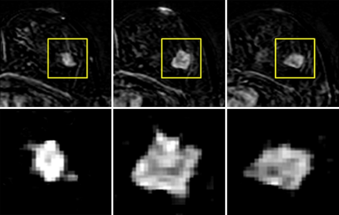
Fig. 2.
The magnified view of the tumour ROI drawn from three slices of the mass lesion shown in Fig. 1. The subtraction image at 1-min after contrast injection is shown. The in-plane resolution is 1.4 × 1.4 mm, and the slice thickness is 4 mm. There is no visible spiculation, which may be due to the relatively low spatial resolution, as well as any slight motion between pre- and post-contrast images

