Fig. 3.
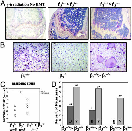
BMT of β3–/– marrow confers protection from osteolytic metastases. (A) (Left) TRAP staining of femur 10 days after 950-rad γ-irradiation in untransplanted control mouse demonstrating fatty marrow devoid of red marrow cells and loss of TRAP+ OCs at growth plate. Recovery of TRAP+ OCs is seen at growth plates of femurs 10 days after BMT of β3+/+ marrow into β3+/+-positive control (Center) or into β3–/– mouse (Right). (B) In vitro TRAP staining of cultured OCs results in multinucleated β3+/+ OCs with well formed actin rings, compared with β3–/– OCs at day 5. Three weeks after BMT with β3+/+ marrow into β3–/– mice restores OC with β3+/+ phenotype (Right). (C) Bleeding times returned to normal range in β3–/– mice 3 weeks after transplantation with β3+/+ marrow. (D) Percentage of transplanted mice with visible bone metastases (B) and visceral metastases (V) 14 days after LV injection of B16 cells. β3+/+>β3+/+ is positive control β3+/+ marrow transplanted into β3+/+ mouse (n = 10). β+/+>β3–/– is β3+/+ marrow transplanted into β3–/– animals (n = 13), demonstrating that β+/+ marrow can restore ability of B16 to induce bone metastases in β3–/– mice. β3–/–>β3+/+ is β3–/– marrow transplanted into β3+/+ mice (n = 7), demonstrating that β3–/– bone marrow can protect WT mice from the bone metastases susceptibility as compared with β3+/+ mice (P = 0.0004 using the Fisher exact t test).

