Fig. 5.
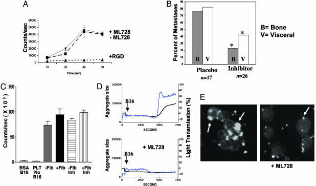
αIIbβ3 inhibitor of platelet aggregation reduces metastases in β3+/+ mice. ML464 is an oral αIIbβ3 antagonist. ML728 is the active metabolite of ML464. (A) B16 cells spreading on fibrinogen-coated surface (♦) were not inhibited by 37.5 μM ML728 (▪) but were completely inhibited by RGD peptide (▴). (B) ML464 (inhibitor) was administered to WT (β3+/+ mice) 30 min before B16 LV injection and then every 12 h for 2.5 days. Placebo in DMSO carrier was also administered by oral gavage. Mice were evaluated 14 days after B16 injection for bone and visceral metastases. Percentage of mice with bone (B) or visceral (V) metastases or placebo-treated mice (n = 17) and ML464 inhibitor-treated mice (n = 26) is shown. *, Metastases were decreased in inhibitor-treated mice compared with placebo-treated mice (P = 0.0013 for bone and P = 0.0125 for visceral using Fisher's exact t test). (C) B16 cells adhered to spread platelets in the presence (+Fib) or absence (–Fib) of fibrinogen, which was not inhibited by ML728 αIIbβ3 inhibitor (Inh). Platelets alone (PLT no B16) and B16 cells on BSA-coated surface (B16 BSA) served as controls. (D) B16 cells added to unactivated platelets induce platelet aggregation as measured in an aggregometer. The arrow represents the addition of B16 cells to platelets. The blue line represents microaggregates, and the black line is total aggregates (micro and large). Addition of 5 μM ML728, an αIIbβ3 antagonist, to stirred platelets before addition of B16 cells completely inhibited platelet aggregation. (E) Calcein-labeled fluorescent mouse platelets adhere to unlabeled B16 tumor cells (arrows) and form aggregates of platelets and tumor cells (Left). Addition of 5 μM ML728 inhibited tumor cell and platelet aggregation/clumping but not platelet–tumor cell adhesion (Right).

