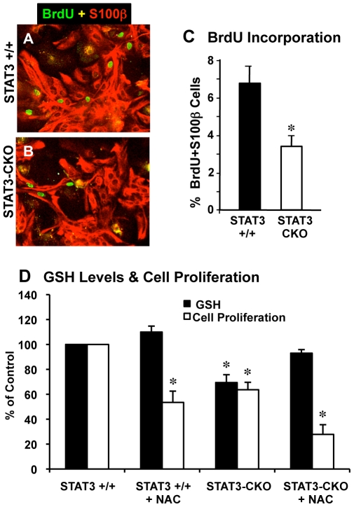
Figure 4. Astrocyte cell proliferation analyzed by BrdU incorporation and propidium iodide fluorescence.
Merged images of double labeling immunofluorescence for S100β and BrdU (A,B) and graph (C) of cell counts show that significantly fewer S100β expressing astrocytes are dividing and labeled with BrdU in STAT3-CKO (B) compared with littermate control (A) cultures (n = 3 per group, * p<0.01 t-test). (D) Cells were cultured to passage 2 over a period of 3 weeks and plated into 96-well plates at a density of 5×10³/well. 0.5 mM N-actetylcysteine (NAC) was added 1, 3, and 5 days after plating. Cell number and GSH assays were performed as described in Materials and Methods. * p<0.01 compared with STAT3 +/+ control using one-way ANOVA with Tukey's post-hoc test.

