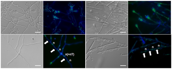
Figure 5. Monokaryotic hyphae, basidia, and basidiospores of F. depauperata.
Hyphae and basidiospores from both strains of F. depauperata appear to be mostly monokaryotic with hyphae that lack clamp connections. Nuclear content was determined by examination under fluorescence light microscopy (color images). Samples were stained with sytox-green to detect nucleic acids (shown in green), and with calcofluor white to visualize the cell walls (shown in blue). DIC images are shown in grey. White arrows indicate cell wall septa, and the letter “n” represents the observed nuclear content. The two left panels are of strain CBS7841, and the two right panels are CBS7855.

