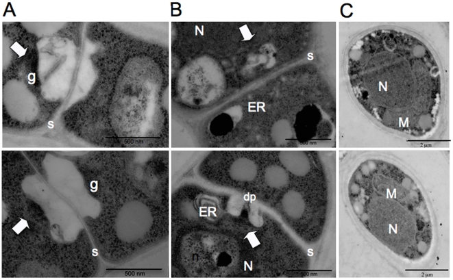
Figure 6. Transmission electron microscopy (TEM) of hyphal septa and basidiospores of F. depauperata.
Panels A and B show different TEM images of the surface view of dolipore septa which is characteristic of basidiomycetes. Panel C shows TEM images from spores. The top row shows TEM images from strain CBS7855 and the bottom row from CBS7841. Arrows indicate electron dense occlusions and vesicles associated with the single septal opening. N denotes the nucleous, M the mitochondria, n the nucleolus, ER the endoplasmic reticulum, dp the dolipore, g the granular dolipore plug, and S the septa. Top panel shows strain CBS7855, and bottom panel shows CBS7841.

