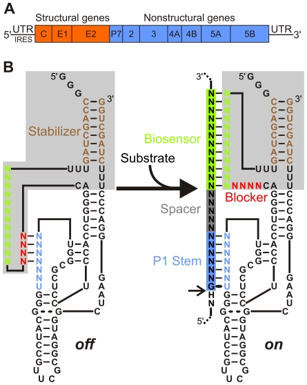
Figure 1. Schematic representations of both the HCV genome and the SOFA-HDV ribozyme.
(A) HCV genomic RNA organization including the structured 5′ and 3′ UTRs. (B) Secondary structure of both the off (left) and on (right) conformations of the SOFA-HDV ribozyme. The SOFA module is highlighted in grey. The P1 domain (ribozyme binding domain), the biosensor, the blocker and the stabilizer domain are indicated blue, green, red and brown, respectively. The spacer region separating the substrate' sequences binding by the P1 and biosensor domains of the ribozyme is indicated in grey. The arrow indicates the cleavage site. All structures have been previously described [29], [30].

