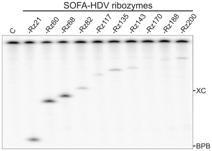
Figure 3. Typical autoradiogram of an 8% polyacrylamide gel performed in order to analyze SOFA-HDV ribozyme cleavage in vitro.
The experiments were performed using 5′-end-labeled HCV transcripts in the presence of an excess of SOFA-HDV ribozyme. The SOFA-HDV ribozymes are identified at the top of the gel. The negative control performed in the absence of ribozyme is indicated by the letter C. The positions of the xylene cyanol (XC) and bromophenol blue (BPB) marker dyes are indicated.

