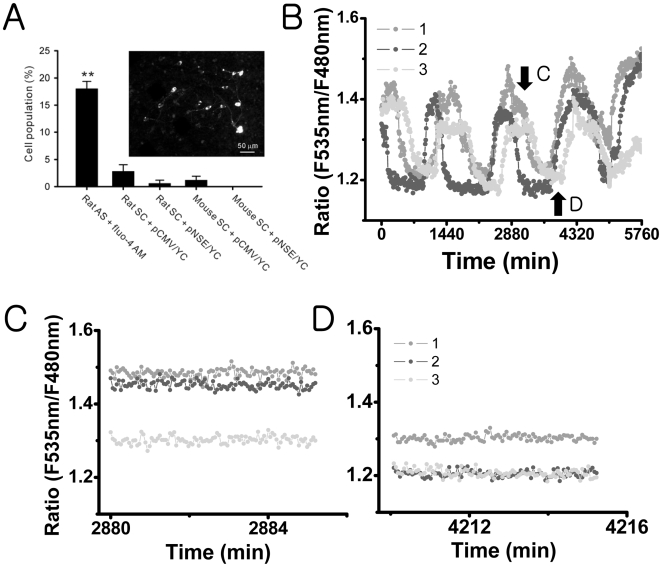
Figure 2. None or fewer Ca2+ spiking activities in cultured SCN cells that express yellow cameleon:
(A) Comparison of cell populations that displayed spontaneous Ca2+ spikes in different SCN preparations. The fluo-4 AM-loaded rat acute slice (rat AS + fluo-4 AM) contains Ca2+ spiking cells in 18% of the total cells (as in Fig. 1). This ratio was significantly larger than that in rat slice cultures (rat SC) or in mouse slice cultures (mouse SC). **P<.01 by one-way ANOVA. The slice cultures that express yellow cameleon with CMV promoter (+pCMV/YC) had slightly larger number of Ca2+ spiking cells in comparison with those that express yellow cameleon with NSE promoter (+pNSE/YC), but the difference was not statistically significant. An example fluorescent image of mouse SC + pNSE/YC is shown on the left (scale bar, 50 µm). (B) Long-term traces (sampling rate at 1 frame per 10 min) of the level of [Ca2+]c in three different SCN neurons in the mouse SC + pNSE/YC represent robust circadian oscillations. The imaging done at the higher sampling rate (1 frame per 3 seconds) was taken at the approximate plateau (C) and trough (D) of the circadian [Ca2+]c rhythms, with no [Ca2+]c spiking activities being detected in these SCN neurons.

