Figure 1.

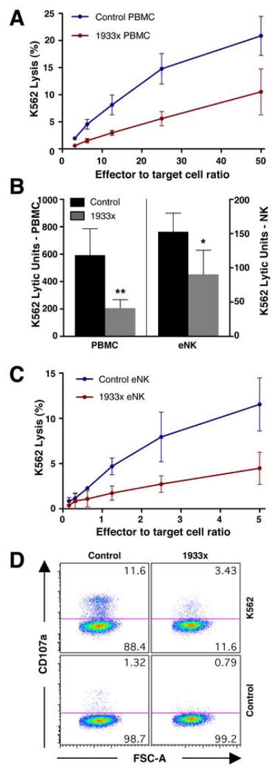
Effect of myosin IIA 1933x mutation on cytotoxicity and perforin localization at the IS. (A) Cytotoxicity of control donor (blue) and myosin IIA 1933x patient (red) PBMCs against K562 target cells. Mean ±SD specific K562 51Cr-release from 3 separate control donors and patients is shown. Individual experiments were performed in triplicate and averages of these were used to calculate means of the patients or controls ± SD. (B) Mean K562 lytic units ±SD of PBMCs and eNK cells from 4 control donors (black) and myosin IIA 1933x patients (gray). Patient and control means were compared using the exact Wilcoxon-Mann-Whitney test: **p=0.014 for PBMCs and *p=0.05 for eNK cells. (C) Cytotoxicity of control donor (blue) and myosin IIA 1933x patient (red) eNK cells against K562 target cells. Mean ±SD specific 51Cr-release from 3 separate control donors and patients is shown. (D) Flow cytometric analysis depicting CD107a exposure in CD3−CD4−CD56+ cells from control donor and myosin IIA 1933x patient PBMC populations mixed with DMSO or K562 cells.
