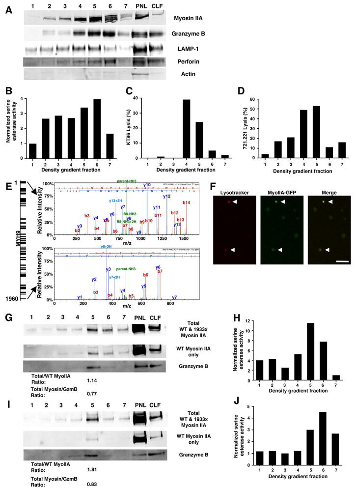
Figure 5.

Evaluation of myosin IIA in isolated NK cell lytic granules. Fractions from density gradient separation of lytic granules from YTS (A–E), YTS-Myosin IIA-GFP (F), control NK (G–H), and 1933X NK (I–J) cells. (A, G, I) Myosin IIA, granzyme B, LAMP-1, perforin, and actin Western blot of density gradient fractions, from least (1) to most dense (7) as well as the post-nuclear lysate (PNL) and crude lysosomal fraction (CLF) generated in preparing the starting material for the density gradient. In (G, I), anti-myosin IIA antibodies with specificities for both wild type and 1933x myosin IIA (top), or only wild-type myosin IIA (middle). Densitometric ratios of total to wild-type epitope recognizing antibody signal, as well as total myosin IIA to granzyme B, are shown below the Western blots. (B, H, J) Serine esterase activity of YTS (B), control NK (H), and 1933x NK cell (J) density gradient fractions. (C–D) Cytotoxicity of KT86 (C) and 721.221 (D) target cells mediated by YTS density gradient fractions. (E) Mass spectrometric analysis of peptides identified in a density gradient fraction from YTS cells enriched in myosin IIA and granzyme B, identifying MYH9 in the bands demonstrated in the Western blots. The diagram (left) depicts peptide coverage of MYH9, and the mass spectra (right) represent the most N-terminal and most C-terminal peptides identified. (F) Confocal fluorescent images of isolated lytic granules from YTS-Myosin IIA-GFP cells (gradient fractions 5 and 6) loaded with Lysotracker Red. Presumed lytic granules (arrows) demonstrated both Lysotracker (red) and myosin IIA-GFP (green) fluorescence. Scale bar = 4 μm.
