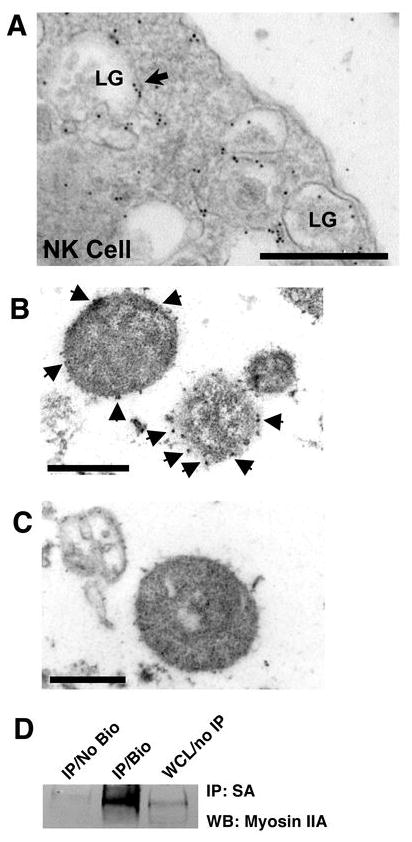
Figure 6.

Ultrastructural localization of myosin IIA in NK cells and isolated lytic granules. (A) Electron micrograph of an unconjugated eNK cell stained with rabbit anti-myosin IIA Ab followed by gold-conjugated anti-rabbit Ab. LG = lytic granule. Arrows indicate areas containing gold particles. Scale bar = 500 nm. (B) Electron micrograph of isolated lytic granules from YTS cells stained with rabbit anti-myosin IIA Ab followed by gold-conjugated anti-rabbit Ab. Arrows indicate areas containing gold particles. Scale bar = 500 nm. (C) Electron micrograph of isolated lytic granules from YTS cells stained with gold-conjugated anti-rabbit Ab alone. Scale bar = 500 nm. Arrows indicate gold particles. (D) Western blot of streptavidin-agarose immunoprecipitates from unlabeled lytic granules (No Bio) and surface-biotinylated lytic granules (Bio), and whole cell lysate (WCL) from YTS cells.
