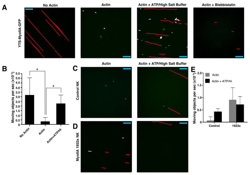
Figure 8.

Functional association of lytic granules with F-actin and effects of ATP and myosin IIA 1933x. Isolated lytic granules were added to flow chambers containing biotinylated BSA/streptavidin (BSA/SA) without actin, with actin and physiologic salt buffer, or with actin and ATP-containing high-salt buffer, or with actin using blebbistatin-treated granules. Granules from YTS-MyoIIA-GFP (A), control NK (C), and 1933x NK (D) cells were drawn through the chamber and allowed to flow through or adhere for 1–2 min before imaging with a confocal microscope at approx. 5 time points per second. From any single culture propagated NK cell preparation the quantity of lytic granules obtained was sufficient for flow through only two chambers. Images were analyzed using the object tracking function in Volocity software and represent an overlay of 12 to 45 seconds. Individual moving objects which had displacement greater than 5 μm (red) and displacement less than 5 μm (white) are indicated. Scale bar = 40 μm. Some images are rotated from their original orientation, shown in Supplementary Videos 4A–6B. (B) Number of moving objects per second ±SD in 3–5 independent sequences of isolated YTS-Myosin IIA-GFP lytic granules added to flow chambers. *= p<0.05.
