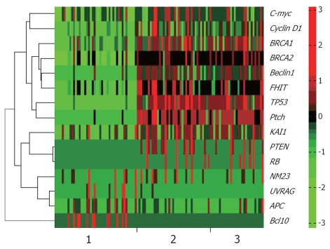
Figure 1.
Gene expression levels of three different tissues. 1: Normal tissues; 2: Well differentiated gastric adenocarcinoma (GA); 3: Poorly differentiated GA. X-axis represents samples, and each unit represents a gene expression level of the corresponding sample. Y-axis represents genes, whose name marked at the right side of the figure. Green color indicates the low-expression level of corresponding gene, and red represents over-expression.

