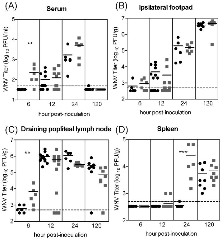
Figure 1.
WNV titers did not differ substantially in mice inoculated with 105 PFU of WNVC6/36 or WNVBHK except at the earliest time points. Mice were inoculated subcutaneously with 105 PFU of WNVC6/36 (●) or WNVBHK ( ) in the left rear footpad. Tissues were harvested at various times post-inoculation, and plaque assays were performed to determine the viral load in (A) serum, (B) ipsilateral footpad (inoculation site), (C) draining popliteal lymph node, and (D) spleen. Data from two independent experiments are shown. Solid lines indicate the geometric mean for each group of 6–12 mice. Dashed lines represent the limit of detection (LOD), and data points below the line are < LOD. Asterisks denote significant difference between groups (Mann-Whitney test: ** p <0.05; ***p <0.005).

