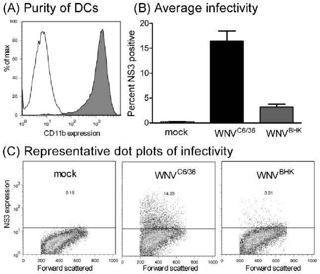
Figure 5.
The percentage of primary dendritic cells infected by WNVC6/36 was greater than the percentage infected by WNVBHK. (A) After 7 days in culture, primary immature DCs were incubated with PE-conjugated anti-CD11b (grey, filled) or isotype control (black, unfilled) prior to infection with WNV. (B and C) Primary DCs were inoculated with diluent (mock) or with WNVC6/36 or WNVBHK at MOI of 50. Cells were harvested at 48 hpi, and incubated with Alexa fluor 647-conjugated antibodies against NS3. All samples were fixed overnight at 4°C and then analyzed by flow cytometry. (B) Average ± standard error from 2 independent experiments and (C) representative dot plots are shown.

