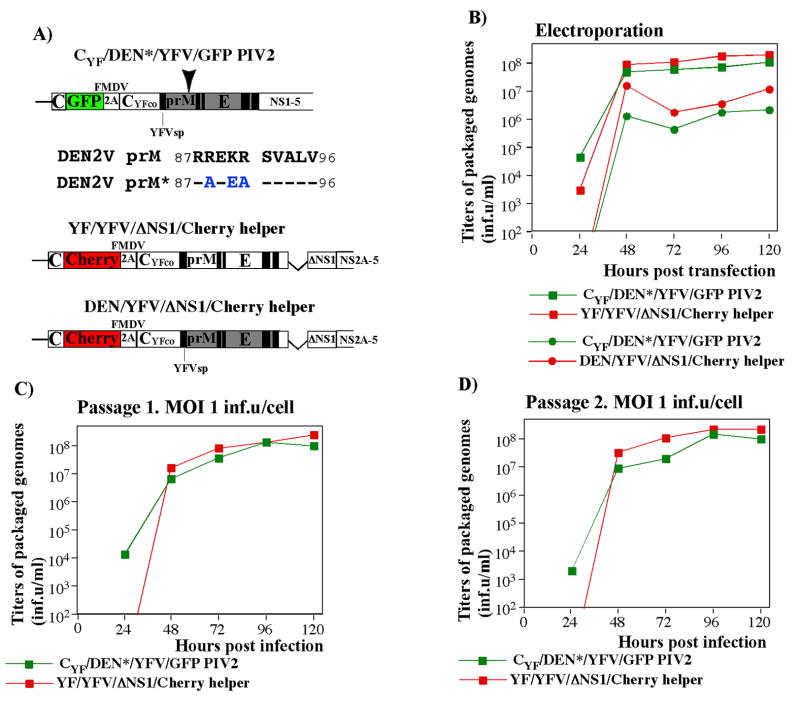
FIG. 4.
Replication of chimeric PIV2 with mutated furin cleavage site in BHK-21 cells. (A) The schematic representation of CYF/DEN*/YFV/GFP PIV2 genome. The position of mutations in prM cleavage site is indicated by arrow. Identical amino acids in the prM alignment are denoted by dashes. The introduced mutations are indicated by blue color. YFV-specific sequences in PIV2 and helper genomes are indicated by open boxes. DEN2V-specific sequences are indicated by filled, grey boxes. FMDV 2A indicates position of FMDV 2A protease. (B) BHK-21 cells were electroporated by the in vitro-synthesized PIV2 and helper RNAs (see Materials and Methods for details). Cells were seeded into 100-mm dishes in 10 ml of media and incubated at 37°C. (C and D) BHK-21 cells were infected by the samples harvested at the previous passage at 96 h post transfection or infection, at an MOI of 1 inf.u/cell. Cells were seeded into 100-mm dishes in 10 ml of media and incubated at 37°C. At the indicated time points (B–D), media were replaced and titers of packaged PIV2 and helper genomes were measured by infecting BHK-21 cells, containing VEErep/Pac-2A-NS1 replicon, by different dilutions of the samples and evaluating the numbers of GFP- and Cherry-positive cells.

