
Fig. 6.
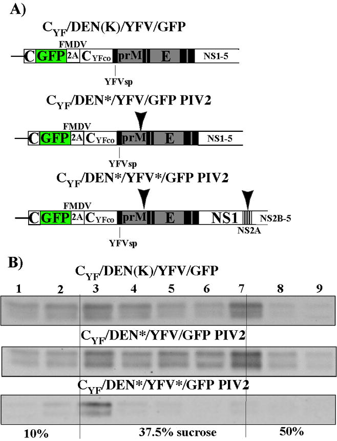
Analysis of viral and subviral particles released from the cells transfected with viral and PIV2 RNAs. (A) Schematic representation of the RNAs used in the experiments. Arrows indicate positions of the mutations introduced into prM and NS2A. (B) Distribution of viral and subviral particles in the discontinuous sucrose gradients. Transfected cells were seeded into 150-mm dishes and, after 24 h incubation at 37°C, media were replaced to serum-free VP-SF medium. After an additional 24 h-long incubation, 20 ml samples of collected media were clarified by low-speed centrifugation and particles were pelleted by ultracentrifugation through the cushion of 10% sucrose in SW-28 rotor (1h, 25, 000 rpm, 4°C). Pellets were suspended in PBS, loaded onto the discontinuous sucrose gradients (see Materials and Methods for details), and centrifugation was performed at 50,000 rpm for 4 h at 4°C in SW-55 rotor of Beckman Optima L-90K ultracentrifuge. Gradients were fractionated, aliquots were diluted with PBS and viruses and VLPs were pelleted in the Beckman TLA-55 rotor. Pellets were dissolved in SDS-containing protein loading buffer having no β-mercaptoethanol. Samples were analyzed by Western blotting using D1-4G2 monoclonal antibody. Secondary goat anti-mouse antibodies were labeled with IRDye 800. Membranes were analyzed on LI-COR imager.