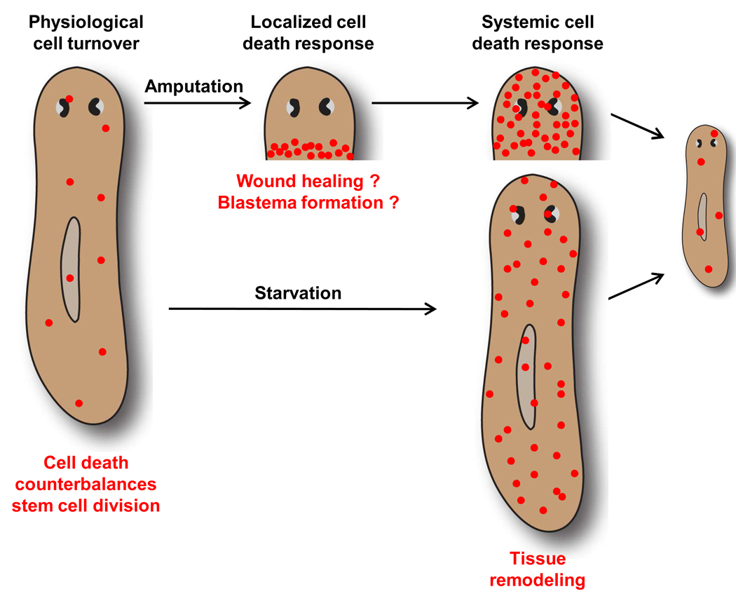
Fig. 7.
Model of apoptosis functions in planarian tissue remodeling. Amputation leads to 2 distinct waves of apoptosis (red circles) in regenerating fragments. The initial localized increase in cell death at 1 to 4 hours post-amputation might promote wound healing and/or blastema formation. We propose that the later systemic increase in cell death represents a key component of the tissue remodeling process that restores scale and proportion and integrates new and preexisting tissues. In this case, remodeling results in a reduction in the size of the photoreceptors, lengthening and narrowing of the entire animal, and integration of the newly formed pharynx. The systemic cell death response is also induced in intact animals by prolonged starvation, contributing to a reduction in the animal’s overall size.

