


Figure 2.
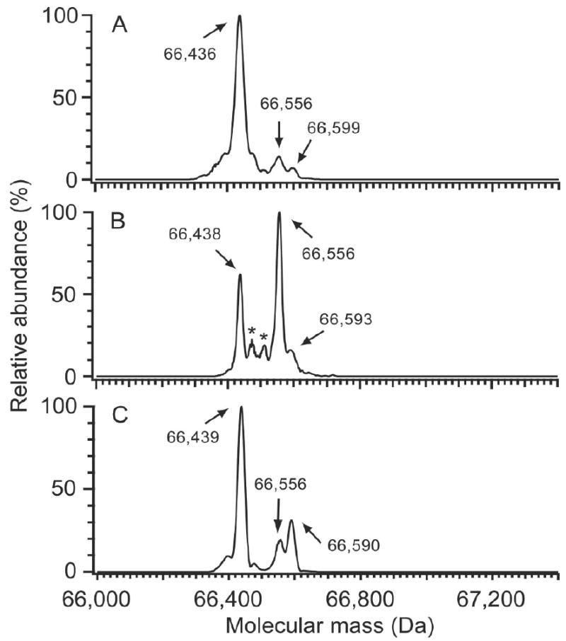
Deconvoluted ESI mass spectra showing heterogeneity of HSA from various sources. (A) Freshly isolated HSA; (B) commercial HSA, and (C) HSA stored at -80°C for 11 years. Mercaptalbumin (66,436 Da) is the most abundant species in fresh HSA (A), with minor peaks at 66,556 Da and 66,599 Da, corresponding to cysteinylation and glycosylation, respectively. Commercial HSA (B) was the most highly oxidized of the three HSA preparations. The most abundant modification was from cysteinylated (66,556 Da), with peaks at 66,473 and 66,593 Da consistent with the addition of NO and glucose, respectively. Archived HSA (C) displayed elevated cysteinylation (66,556 Da) and glycosylation (66,590 Da), and also contained an unknown abundant modification at 66,496 (+60) Da. The modification at 66,496 Da appears to be an irreversible modification at Cys34. The asterisks in (B) denote masses of 66,473 Da and 66,512 Da. (A representative LC-MS total ion chromatogram for the freshly isolated HSA sample is provided in Figure 1A. The raw mass spectrum of the HSA peak from the freshly isolated HSA sample is shown in Figure 1B.)