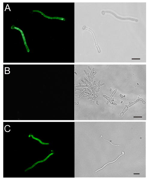
Fig. 6. Indirect immunofluorescence assay (IFA) of wild-type and Δspt6/Δspt6 mutant probed with scFv5.
Wild-type (Panel A), Δspt6/Δspt6 mutant (Panel B), and SPT6 reintegrant cells (Panel C) were induced to form germ tubes and incubated with scFv5 followed by an appropriate fluorochrome labeled secondary antibody. Fluorescence and phase contrast photomicrographs of the same representative microscopic fields are depicted. The typical hyphae-specific binding of scFv5 to wild-type is seen, whereas no binding is detected to the mutant. The reintegrant strain showed binding with a wild-type pattern. Bar – 10 μm.

