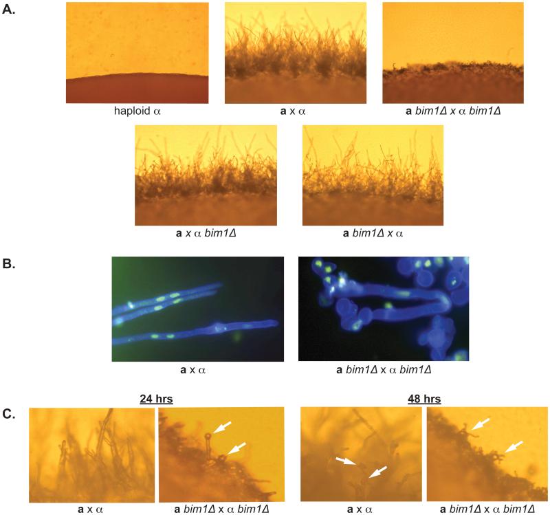
Figure 3.
A. bim1Δ strains show defects during sexual development. Panels show the edges of crosses after 48 hours on V8 medium at 200X magnification. B. Panels show filaments from wild type and mutant crosses stained with calcofluor white (cell wall stain - blue) and Sytox green (nuclear stain) at 1000X magnification. C. Basidia and spore formation occur earlier in bim1Δ strains. Left hand panels show the edge of wild type and bim1Δ crosses taken at 400X magnification after 24 hrs on V8 agar. Right hand panels show the same crosses after 48 hours on V8 agar. White arrows point to basidia and spores.

