
Figure 6.
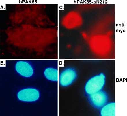
Cell and nuclear changes in cells microinjected with active hPAK65. REF52 cells were microinjected with plasmids encoding GFP and myc-tagged full-length hPAK65 (A and B), and hPAK65-ΔN212 (C and D). Sixteen hours after injection, cells were fixed and stained with immunofluorescent reagents as described in Methods. Pictures were taken from the same field by using different filters. A and C show PAK-expressing cells stained by anti-myc antibody. B and D show nuclei stained by DAPI. Nearly all cells injected with hPAK65-ΔN212 expressed the injected plasmids as evaluated by anti-myc staining (92/96), and greater than 80% of them rounded up (76/92). Injected plasmids were also expressed in most of the cells injected with full-length hPAK65 (114/118); whereas nearly no cell expressing inactive PAK showed any morphological changes (5/114). Similar results have been obtained from three independent experiments.