
Figure 7.
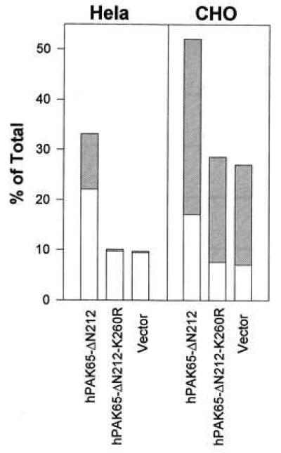
Induction of apoptosis by transfection of active hPAK65 in Hela and CHO cells. The indicated plasmids were cotransfected with surface-expressed CD20 into Hela and CHO cells. Forty hours after transfection, cells, including detached cells, were collected and stained with PE-conjugated CD20, FITC-conjugated annexin V, and PI. Open bars show the percentages of cells in early phase of apoptosis, which are annexin V-positive and PI-negative; hatched bars show the percentages of cells in late phase of apoptosis, which are annexin V- and PI-double-positive. Data represent means of three individual experiments. A similar level of protein expression among all constructs was confirmed by immunoblot analysis (data not shown).