
Figure 8.
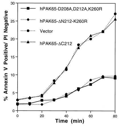
Apoptosis delayed by transfection of dominant-negative hPAK65 in CHO cells stably expressing a CD4-Fas chimera. Forty hours after transfection, cells were collected and stained with PE-conjugated CD20 and CD4. Apoptosis was initiated by adding anti-mouse IgG + IgM, and cells were incubated at 37°C. Aliquots were taken at times indicated, stained with FITC-conjugated annexin V and PI for 20 min at room temperature, and analyzed immediately by flow cytometry.