Figure 6.
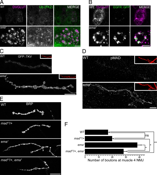
Up-regulated BMP signaling promotes synaptic overgrowth in the ema mutant. (A and B) Endolysosomal trafficking of membrane receptor cargos in motoneurons. Representative confocal images of motoneuron cell bodies labeled for (A) ubiquitinated proteins by a monoclonal α-FK2 antibody (green) or (B) EGFR::GFP fusion protein (green) and for DVGLUT protein (magenta). Bar, 5 µm. (C and D) Distribution of BMP signaling components at the NMJs. Representative confocal images of muscle 4 type 1b NMJs labeled for (C) neuronally expressed GFP::TKV fusion protein and (D) endogenous pMAD protein. In insets, the NMJs are visualized by the neuronal membrane marker HRP (red). Bar, 20 µm. (E and F) A mad mutation dominantly suppresses the synaptic overgrowth in ema. (E) Representative confocal images of wild type (WT), a heterozygous mad mutant (mad12/+), ema1, and mad12/+, ema1 NMJs labeled for the presynaptic protein BRP. Bar, 20 µm. (F) Quantification of number of synaptic boutons at the muscle 4 NMJs for the genotypes in E. n > 10 for all genotypes. Data represent mean ± SEM. ANOVA analysis. *, P < 0.05; **, P < 0.01; ns, not significant.

