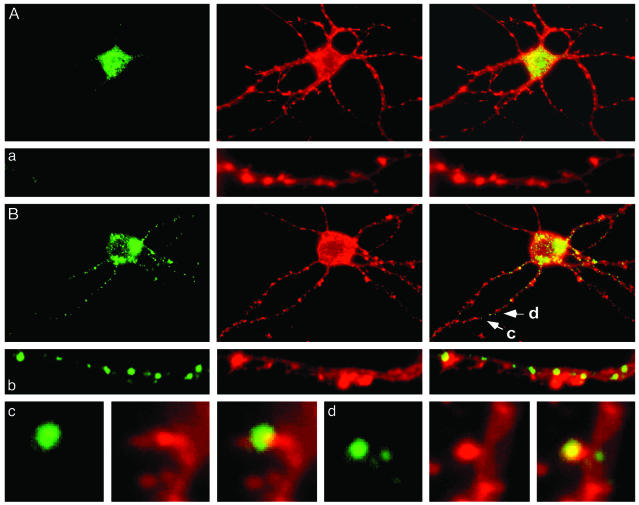
Fig. 5.
Dendrite-wide induction of Thr-389-P p70S6K in primary hippocampal neurons. (A) Thr-389-P p70S6K was found predominantly in the soma of primary hippocampal neurons (21 days in vitro) (labeling for Thr-389-P p70S6K with a phosphorylation state-specific rabbit polyclonal antibody is shown in green (Right), counterstaining for actin with phycoerythrin-conjugated phalloidin in red in the middle, and the overlay in yellow (Left). Similar results were obtained with a mouse monoclonal antibody specific for Thr-389-P p70S6K. A representative dendrite is shown in a. (B) Thr-389-P p70S6K appeared in distinct punctae along dendrites after depolarization with KCl. A representative dendrite is shown in b. In dendrites (c and d), Thr-389-P p70S6K was also present in a subset of dendritic spines as revealed by double labeling for actin filaments (c and d as shown in B). The morphology of Thr-389-P p70S6K-immunoreactive puncta in dendritic shafts was reminiscent of the morphology of hotspots of dendritic translation recently demonstrated by Aakalu et al. and Job and Eberwine (3, 4) in similarly cultured primary hippocampal neurons.

