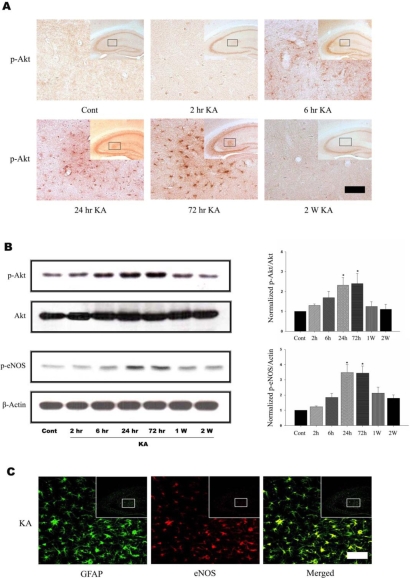
Fig. 1.
KA induces Akt and eNOS phosphorylation in mouse hippocampus. (A) Time course of KA-induced Akt phosphorylation in astrocytes. Increased p-Akt immunoreactivity was observed predominantly in astrocytes in the hippocampus 6 h after KA treatment and maintained up to 72 h. Inset at right upper corner shows the position of the enlarged image of CA3 region. (B) Representative immunoblots (left) and quantitative analysis (right) of Akt and eNOS phosphorylation. The levels of phosphorylation of Akt at Ser473 and eNOS at Ser1177 were measured after KA injection. Phosphorylation of Akt and eNOS increased up to 3 days, and then normalized 2 weeks after KA injection. (C) Confocal image of astrocytic eNOS in the hippocampus. Double immunofluorescence staining was carried out with antibodies to eNOS and GFAP, an astrocytic marker. eNOS immunoreactivity appeared to co-localize with that of GFAP. Data represent three independent experiments and were expressed mean±SD. *p<0.05 indicate statistically significant difference from control group. Scale bar: 50µm.

