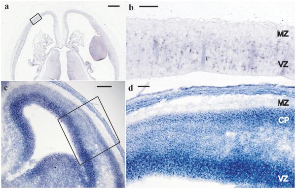
Figure 5. PNKP in situ hybridization.
In situ hybridization of Carnegie Stage 22 human embryos (~54 postovulatory days) with anti-sense probe to human PNKP (a). Sense strand (not shown) showed no specific hybridization. Higher magnification image of developing cerebral cortex boxed area in (a) is shown in (b). Ventricular zone (VZ), containing proliferating cells, shows PNKP mRNA expression while the cell-sparse marginal zone (MZ) has no staining. Mouse E14 cerebral cortex (c) with high magnification of boxed region shown in (d) shows a similar staining pattern with high expression within the proliferating VZ and lower but maintained expression within differentiated neurons of the cortical plate (CP). (a) and (b) are in the transverse plane and (c) and (d) are coronal. Scale bars, (a) 1 mm, (b) 100 μm, (c) 150 μm, (d) 75 μm.

