Fig. 1.
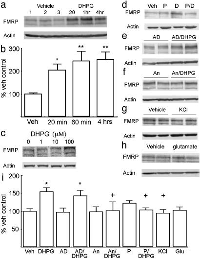
FMRP expression is up-regulated in cortical cultures in response to activation of type-I mGluRs. (a) A representative immunoblot of FMRP (Upper) and β-actin (Lower) expression in cultured neurons treated with vehicle or 100 μM DHPG for the times shown. (b) Summary of multiple experiments (more than five in all cases) evaluating the effect of DHPG on FMRP expression. (c) Representative immunoblot showing the effects of different concentrations of DHPG on FMRP expression at 20 min. Lanes from left to right are 0, 1, 10, and 100 μM.(d) Effect of mGluR antagonist on DHPG-mediated FMRP expression. Immunoblot of FMRP after vehicle alone (Veh), the type-I mGluR antagonist PHCCC (P), 20 min of DHPG alone (D), or PHCCC and DHPG together (P/D) is shown. (e and f) Representative immunoblots showing FMRP expression after treatment with actinomycin D (AD) (e) or anisomycin (An) (f). After a 10-min pretreatment, cells were treated with either vehicle (AD) or DHPG (AD/DHPG) for 20 min. (g) FMRP after 20 min of 50 mM KCl. (h) FMRP after 20 min of 50 μM glutamate. (i) Summary of experiments shown in d-h on the expression of FMRP in cultured neurons. Throughout figure, y axes are percentage vehicle controls, and error bars represent SEM. **, P < 0.0001 versus control; *, P < 0.05 versus control; +, P < 0.05 versus 20-min DHPG treatment alone.

