Fig. 1.
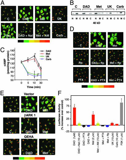
Activation of DOR or CB1 promotes PKA Cα translocation and CRE-Luc expression in NG cells. (A) Cα translocation detected by immunostaining. NG cells were incubated with or without 1 μM DADLE (DAD), 2 μM Met, 10 μM UK, or 10 μM Carb or forskolin (FSK) (1 μM) for 10 min. Where indicated, cells were preincubated either with the DOR antagonist Nal (10 μM) or the CB1 antagonist AM (10 μM) for 30 min. Data represent at least three experiments. Staining intensity is indicated by the color bar (red indicates highest concentration) (scale bar, 10 μm; magnification, ×400). (B)Cα translocation detected by Western blots of nuclear (N), membrane (M), and cytosolic (C) fractions from treated cells. (C) Time course of cAMP production. Cells were incubated with or without 1 μM DADLE, 2 μM Met, 10 μM UK, or 10 μM Carb for the indicated times. cAMP was measured by RIA (17). Data are the mean ± SEM of four experiments. *, P < 0.05 compared with time 0 (one-way analysis of variance and Dunnett's test). cAMP levels in the absence of drugs did not change during the experiment. (D) Rp and PTX inhibit PKA Cα translocation. Cells were pretreated with 20 μM Rp for 1.5 h or PTX (50 ng/ml) overnight before incubation with DADLE or Met as in A. (E) βγ dimers are required for PKA Cα translocation. Cells were transfected with the βγ inhibitors Ad5βARK1 or Ad5QEHA or Ad5 vector control and incubated with or without 1 μM DADLE or 2 μM Met. (F) Activation of DOR or CB1 induces CRE-Luc expression. Cells were transiently transfected with a CRE-Luc construct, preincubated with buffer or Nal, AM, Rp, or PTX as above and then treated for 10 min with or without DADLE, Met, or forskolin. Luc was assayed 5 h after drug treatment. Data are the mean ± SEM of at least three experiments. *, P < 0.01 compared with control (one-way analysis of variance and Dunnett's test).

