Figure 6.
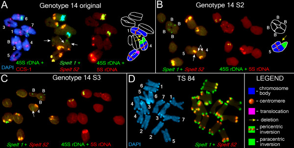
Fluorescence in situ hybridization (FISH) and differential staining with 4',6-diamidino-2-phenylindole (DAPI) on somatic and meiotic chromosomes of Aegilops speltoides (part 2). (a) From left to right: DAPI and FISH with the CCS-1 and 45S rDNA on the meiotic chromosomes of the original G14 plant; FISH with Spelt 52 and Spelt 1. The clusters of Spelt 1 that mark a homozygous paracentric inversion on chromosome 4, and a Spelt 1 cluster on the B chromosome are arrowed. FISH with 5S rDNA and 45S rDNA on the same chromosomes. A scheme of the main chromosomal rearrangements. (b) FISH with Spelt 52 and Spelt 1 on the meiotic chromosomes of the G14 S2 plant (left). The clusters of Spelt 1 that mark a homozygous paracentric inversion on chromosome 4 and cluster of Spelt 52 that marks a heterozygous inversion on the chromosome 5 are arrowed. FISH with 5S rDNA and 45S rDNA (middle). B chromosomes carry 5S rDNA clusters in both arms. The scheme of the main chromosomal rearrangements (right). (c) FISH with Spelt 52 and Spelt 1 on the meiotic chromosomes of the G14 S3 plant (left). Homozygous paracentric inversion on chromosome 4 is arrowed. FISH with 5S rDNA and 45S rDNA on the same chromosomes (right). (d) Somatic chromosomes of TS 84, staining with DAPI (left). FISH with Spelt 52 and Spelt 1 on the same chromosomes (right). The DNA probes and staining: (a-d) 5S rDNA, Spelt 52 and cereal centromere-specific sequence 1 (CCS-1) in red; 45S rDNA and Spelt 1 in green; differential staining with DAPI in blue; (a) 5S rDNA (yellow) and 45S rDNA (blue) in pseudocolors.

